Newest technological advance in recent history
Panoramic dental x-ray uses a very small dose of ionizing radiation to capture the entire mouth in one image. It is commonly performed by dentists and oral surgeons in everyday practice and may be used to plan treatment for dentures, braces, extractions and implants.
What is Panoramic X-Ray?
Panoramic radiography, also called panoramic x-ray, is a two-dimensional (2-D) dental x-ray examination that captures the entire mouth in a single image, including the teeth, upper and lower jaws, surrounding structures and tissues.
The jaw is a curved structure similar to that of a horseshoe. However, the panoramic x-ray produces a flat image of the curved structure. It usually provides details of the bones and teeth.
An x-ray (radiograph) is a noninvasive medical test that helps physicians diagnose and treat medical conditions. Imaging with x-rays involves exposing a part of the body to a small dose of ionizing radiation to produce pictures of the inside of the body. X-rays are the oldest and most frequently used form of medical imaging.
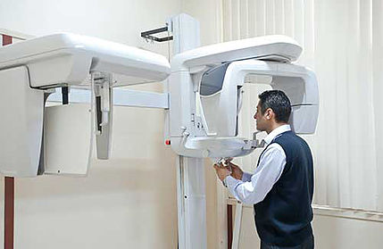
Unlike a traditional intraoral x-ray where the film/x-ray detector is placed inside of the mouth, the film for a panoramic x-ray is contained inside of the machine.
This exam requires little to no special preparation. Tell your doctor if there is a possibility you are pregnant. Remove any jewelry, eye glasses or metal objects that might interfere with the x-ray images. You will be asked to wear a lead apron to protect the rest of your body from any radiation exposure.